*Ado Oculus Imaging Classes
CT image of the right ventricle
 |
 |
 |
 |
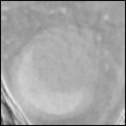
First:Test image. It is an ultra-fast CT image of the right ventricle of the canine.
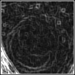
Second: Gradient image.
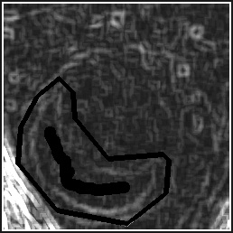
Third: Interactively imposed markers (really dark pixel areas).
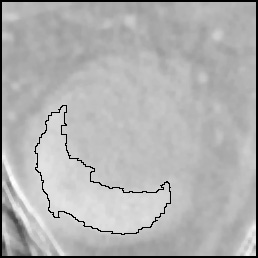
Fourth: Meyer's watershed transform. The water coming from the markers floods all the irrelevant catchment basins except the highest-crest line around the object marker. The segmentation result looks visually sensible.1. How can the markers be automatically detected? Questions:
2. What could be the final wateshed lines if the object marker is a circle instead of an elongated bar?