Common Names: Threshold, Density slicing
In many vision applications, it is useful to be able to separate out
the regions of the image corresponding to objects in which we are
interested, from the regions of the image that correspond to
background. Thresholding often provides an easy and convenient way to
perform this segmentation on the basis of the different intensities or
colors in the foreground and background regions of an image.
In addition, it is often useful to be able to see what areas of an image consist of pixels whose values lie within a specified range, or band of intensities (or colors). Thresholding can be used for this as well.
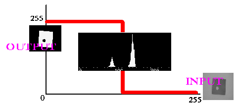
The input to a thresholding operation is typically a grayscale or color image. In the simplest implementation, the output is a binary image representing the segmentation. Black pixels correspond to background and white pixels correspond to foreground (or vice versa). In simple implementations, the segmentation is determined by a single parameter known as the intensity threshold. In a single pass, each pixel in the image is compared with this threshold. If the pixel's intensity is higher than the threshold, the pixel is set to, say, white in the output. If it is less than the threshold, it is set to black.
In more sophisticated implementations, multiple thresholds can be specified, so that a band of intensity values can be set to white while everything else is set to black. For color or multi-spectral images, it may be possible to set different thresholds for each color channel, and so select just those pixels within a specified cuboid in RGB space. Another common variant is to set to black all those pixels corresponding to background, but leave foreground pixels at their original color/intensity (as opposed to forcing them to white), so that that information is not lost.
Not all images can be neatly segmented into foreground and background using simple thresholding. Whether or not an image can be correctly segmented this way can be determined by looking at an intensity histogram of the image. We will consider just a grayscale histogram here, but the extension to color is trivial.
If it is possible to separate out the foreground of an image on the basis of pixel intensity, then the intensity of pixels within foreground objects must be distinctly different from the intensity of pixels within the background. In this case, we expect to see a distinct peak in the histogram corresponding to foreground objects such that thresholds can be chosen to isolate this peak accordingly. If such a peak does not exist, then it is unlikely that simple thresholding will produce a good segmentation. In this case, adaptive thresholding may be a better answer.
Figure 1 shows some typical histograms along with suitable choices of threshold.
Figure 1 A) shows a classic bi-modal intensity distribution. This image can be successfully segmented using a single threshold T1. B) is slightly more complicated. Here we suppose the central peak represents the objects we are interested in and so threshold segmentation requires two thresholds: T1 and T2. In C), the two peaks of a bi-modal distribution have run together and so it is almost certainly not possible to successfully segment this image using a single global threshold
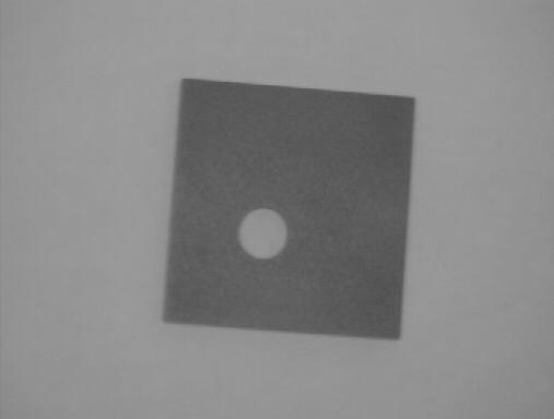
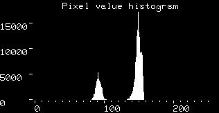
The histogram for image
is
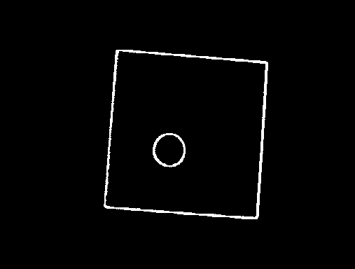
This shows a nice bi-modal distribution --- the lower peak represents the object and the higher one represents the background. The picture can be segmented using a single threshold at a pixel intensity value of 120. The result is shown in
The histogram for image
is
Due to the severe illumination gradient across the scene, the peaks corresponding to foreground and background have run together and so simple thresholding does not give good results. Images
and
show the resulting bad segmentations for single threshold values of 80 and 120 respectively (reasonable results can be achieved by using adaptive thresholding on this image).
Thresholding is also used to filter the output of or input to other operators. For instance, in the former case, an edge detector like Sobel will highlight regions of the image that have high spatial gradients. If we are only interested in gradients above a certain value (i.e. sharp edges), then thresholding can be used to just select the strongest edges and set everything else to black. As an example,
was obtained by first applying the Sobel operator to
to produce
and then thresholding this using a threshold value of 60.
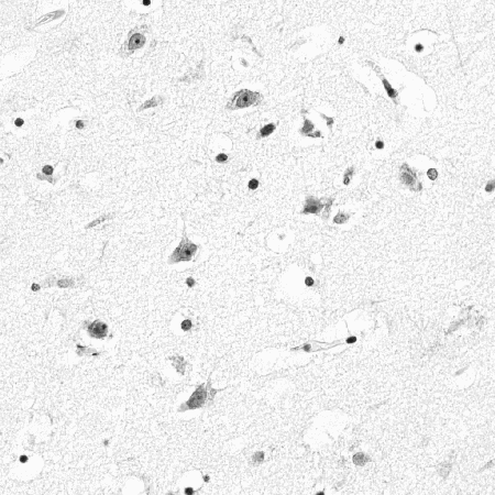
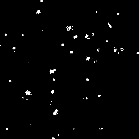
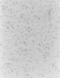
Thresholding can be used as preprocessing to extract an interesting subset of image structures which will then be passed along to another operator in an image processing chain. For example, image
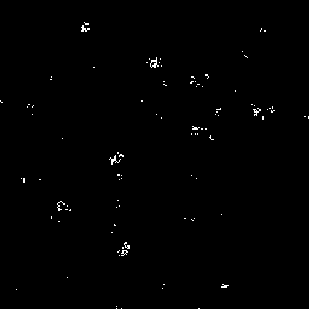
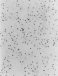
shows a slice of brain tissue containing nervous cells (i.e. the large gray blobs, with darker circular nuclei in the middle) and glia cells (i.e. the isolated, small, black circles). We can threshold this image so as to map all pixel values between 0 and 150 in the original image to foreground (i.e. 255) values in the binary image, and leave the rest to go to background, as in
The resultant image can then be connected-components-labeled in order to count the total number of cells in the original image, as in
If we
wanted to know how many nerve cells there are in the original image,
we might try applying a double threshold in order to select out just
the pixels which correspond to nerve cells (and therefore have middle
level grayscale intensities) in the original image. (In remote sensing
and medical terminology, such thresholding is usually called
density slicing.) Applying a threshold band of 130 - 150 yields
While most of the foreground of the resulting image corresponds to nerve cells, the foreground features are so disconnected (because nerve cell nuclei map to background intensity values along with the glia cells) that we cannot apply connected components labeling. Alternatively, we might obtain a better assessment of the number of nerve cells by investigating some attributes (e.g. size, as measured by a distance transform) of the binary image containing both whole nerve cells and glia. In reality, sophisticated modeling and/or pattern matching is required to segment such an image.
You can interactively experiment with this operator by clicking here.
and
were each taken on a different day during an experiment which sought to quantify cell death. Investigate the intensity histogram of these images and choose a threshold which allows you to segment out the dead cells. Then use connected components labeling to count the number of dead cells on each day of the experiment.
assume that wheat fields are visible as yellow patches. Construct a set of thresholds for each color channel which allow you to segment out the wheat fields (note, you may need to reset your display).
E. Davies Machine Vision: Theory, Algorithms and Practicalities, Academic Press, 1990, Chap. 4.
R. Gonzalez and R. Woods Digital Image Processing, Addison-Wesley Publishing Company, 1992, Chap. 7.
D. Vernon Machine Vision, Prentice-Hall, 1991, pp 49 - 51, 86 - 89.
Specific information about this operator may be found here.
More general advice about the local HIPR installation is available in the Local Information introductory section.
©2003 R. Fisher, S. Perkins,
A. Walker and E. Wolfart.
![]()